Cookies Policy
The website need some cookies and similar means to function. If you permit us, we will use those means to collect data on your visits for aggregated statistics to improve our service. Find out More
Accept Reject
  • Menu
About

About

My name is Ana Maria Mendonça and I am currently Associate Professor at the Department of Electrical and Computer Engineering (DEEC) of the Faculty of Engineering of the University of Porto (FEUP), where I got my PhD in 1994. I was a researcher at the Institute for Biomedical Engineering (INEB) until 2014, but since 2015 I am a senior researcher at INESC. At INEB, I was a member of the Board of Directors and afterwards President of the Board.

In my management activities in higher education and research, I was a member of the Executive Board of DEEC and more recently Deputy Director of FEUP. At INEB, I was a member of the Institute's Board of Directors, initially as a member and later as President of the Board.

I was an elected member of FEUP's Scientific Council and am currently a member of the school's Pedagogical Council. I was a member of the scientific committee of several academic programmes and, currently, I am the Director of the First Degree and the Master Degree in BioEngineering, of the Biomedical Engineering Master and the Doctoral Programme in Biomedical Engineering.

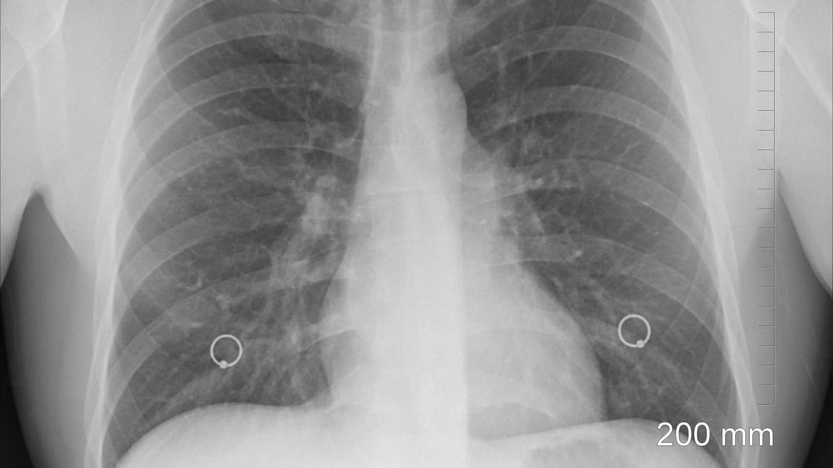
I have been collaborating as a research and also as responsible in several research projects, mostly dedicated to the development of image analysis and classification methodologies aiming at extracting essential information from medical images in order to support the diagnosis process. Past work has been mostly devoted to three main areas: retinal pathologies, lung diseases and genetic disorders, but ongoing work is mainly focused on the development of Computer-Aided Diagnosis systems in Ophthalmology and Radiology.

Interest
Topics
Details

Details

  • Name

    Ana Maria Mendonça
  • Role

    Senior Researcher
  • Since

    01st January 2015
  • Nationality

    Portugal
  • Contacts

    +351222094106
    ana.mendonca@inesctec.pt
005
Publications

2024

STERN: Attention-driven Spatial Transformer Network for abnormality detection in chest X-ray images

Authors
Rocha, J; Pereira, SC; Pedrosa, J; Campilho, A; Mendonça, AM;

Publication
ARTIFICIAL INTELLIGENCE IN MEDICINE

Abstract
Chest X-ray scans are frequently requested to detect the presence of abnormalities, due to their low-cost and non-invasive nature. The interpretation of these images can be automated to prioritize more urgent exams through deep learning models, but the presence of image artifacts, e.g. lettering, often generates a harmful bias in the classifiers and an increase of false positive results. Consequently, healthcare would benefit from a system that selects the thoracic region of interest prior to deciding whether an image is possibly pathologic. The current work tackles this binary classification exercise, in which an image is either normal or abnormal, using an attention-driven and spatially unsupervised Spatial Transformer Network (STERN), that takes advantage of a novel domain-specific loss to better frame the region of interest. Unlike the state of the art, in which this type of networks is usually employed for image alignment, this work proposes a spatial transformer module that is used specifically for attention, as an alternative to the standard object detection models that typically precede the classifier to crop out the region of interest. In sum, the proposed end-to-end architecture dynamically scales and aligns the input images to maximize the classifier's performance, by selecting the thorax with translation and non-isotropic scaling transformations, and thus eliminating artifacts. Additionally, this paper provides an extensive and objective analysis of the selected regions of interest, by proposing a set of mathematical evaluation metrics. The results indicate that the STERN achieves similar results to using YOLO-cropped images, with reduced computational cost and without the need for localization labels. More specifically, the system is able to distinguish abnormal frontal images from the CheXpert dataset, with a mean AUC of 85.67% -a 2.55% improvement vs. the 0.98% improvement achieved by the YOLO-based counterpart in comparison to a standard baseline classifier. At the same time, the STERN approach requires less than 2/3 of the training parameters, while increasing the inference time per batch in less than 2 ms. Code available via GitHub.

2024

Automated image label extraction from radiology reports - A review

Authors
Pereira, SC; Mendonca, AM; Campilho, A; Sousa, P; Lopes, CT;

Publication
ARTIFICIAL INTELLIGENCE IN MEDICINE

Abstract
Machine Learning models need large amounts of annotated data for training. In the field of medical imaging, labeled data is especially difficult to obtain because the annotations have to be performed by qualified physicians. Natural Language Processing (NLP) tools can be applied to radiology reports to extract labels for medical images automatically. Compared to manual labeling, this approach requires smaller annotation efforts and can therefore facilitate the creation of labeled medical image data sets. In this article, we summarize the literature on this topic spanning from 2013 to 2023, starting with a meta-analysis of the included articles, followed by a qualitative and quantitative systematization of the results. Overall, we found four types of studies on the extraction of labels from radiology reports: those describing systems based on symbolic NLP, statistical NLP, neural NLP, and those describing systems combining or comparing two or more of the latter. Despite the large variety of existing approaches, there is still room for further improvement. This work can contribute to the development of new techniques or the improvement of existing ones.

2024

Human versus Artificial Intelligence: Validation of a Deep Learning Model for Retinal Layer and Fluid Segmentation in Optical Coherence Tomography Images from Patients with Age-Related Macular Degeneration

Authors
Miranda, M; Santos-Oliveira, J; Mendonca, AM; Sousa, V; Melo, T; Carneiro, A;

Publication
DIAGNOSTICS

Abstract
Artificial intelligence (AI) models have received considerable attention in recent years for their ability to identify optical coherence tomography (OCT) biomarkers with clinical diagnostic potential and predict disease progression. This study aims to externally validate a deep learning (DL) algorithm by comparing its segmentation of retinal layers and fluid with a gold-standard method for manually adjusting the automatic segmentation of the Heidelberg Spectralis HRA + OCT software Version 6.16.8.0. A total of sixty OCT images of healthy subjects and patients with intermediate and exudative age-related macular degeneration (AMD) were included. A quantitative analysis of the retinal thickness and fluid area was performed, and the discrepancy between these methods was investigated. The results showed a moderate-to-strong correlation between the metrics extracted by both software types, in all the groups, and an overall near-perfect area overlap was observed, except for in the inner segment ellipsoid (ISE) layer. The DL system detected a significant difference in the outer retinal thickness across disease stages and accurately identified fluid in exudative cases. In more diseased eyes, there was significantly more disagreement between these methods. This DL system appears to be a reliable method for accessing important OCT biomarkers in AMD. However, further accuracy testing should be conducted to confirm its validity in real-world settings to ultimately aid ophthalmologists in OCT imaging management and guide timely treatment approaches.

2024

Distribution-based detection of radiographic changes in pneumonia patterns: A COVID-19 case study

Authors
Pereira, SC; Rocha, J; Campilho, A; Mendonça, AM;

Publication
HELIYON

Abstract
Although the classification of chest radiographs has long been an extensively researched topic, interest increased significantly with the onset of the COVID-19 pandemic. Existing results are promising; however, the radiological similarities between COVID-19 and other types of respiratory diseases limit the success of conventional image classification approaches that focus on single instances. This study proposes a novel perspective that conceptualizes COVID-19 pneumonia as a deviation from a normative distribution of typical pneumonia patterns. Using a population- based approach, our approach utilizes distributional anomaly detection. This method diverges from traditional instance-wise approaches by focusing on sets of scans instead of individual images. Using an autoencoder to extract feature representations, we present instance-based and distribution-based assessments of the separability between COVID-positive and COVIDnegative pneumonia radiographs. The results demonstrate that the proposed distribution-based methodology outperforms conventional instance-based techniques in identifying radiographic changes associated with COVID-positive cases. This underscores its potential as an early warning system capable of detecting significant distributional shifts in radiographic data. By continuously monitoring these changes, this approach offers a mechanism for early identification of emerging health trends, potentially signaling the onset of new pandemics and enabling prompt public health responses.

2023

Lightweight multi-scale classification of chest radiographs via size-specific batch normalization

Authors
Pereira, SC; Rocha, J; Campilho, A; Sousa, P; Mendonca, AM;

Publication
COMPUTER METHODS AND PROGRAMS IN BIOMEDICINE

Abstract
Background and Objective: Convolutional neural networks are widely used to detect radiological findings in chest radiographs. Standard architectures are optimized for images of relatively small size (for exam-ple, 224 x 224 pixels), which suffices for most application domains. However, in medical imaging, larger inputs are often necessary to analyze disease patterns. A single scan can display multiple types of radi-ological findings varying greatly in size, and most models do not explicitly account for this. For a given network, whose layers have fixed-size receptive fields, smaller input images result in coarser features, which better characterize larger objects in an image. In contrast, larger inputs result in finer grained features, beneficial for the analysis of smaller objects. By compromising to a single resolution, existing frameworks fail to acknowledge that the ideal input size will not necessarily be the same for classifying every pathology of a scan. The goal of our work is to address this shortcoming by proposing a lightweight framework for multi-scale classification of chest radiographs, where finer and coarser features are com-bined in a parameter-efficient fashion. Methods: We experiment on CheXpert, a large chest X-ray database. A lightweight multi-resolution (224 x 224, 4 48 x 4 48 and 896 x 896 pixels) network is developed based on a Densenet-121 model where batch normalization layers are replaced with the proposed size-specific batch normalization. Each input size undergoes batch normalization with dedicated scale and shift parameters, while the remaining parameters are shared across sizes. Additional external validation of the proposed approach is performed on the VinDr-CXR data set. Results: The proposed approach (AUC 83 . 27 +/- 0 . 17 , 7.1M parameters) outperforms standard single-scale models (AUC 81 . 76 +/- 0 . 18 , 82 . 62 +/- 0 . 11 and 82 . 39 +/- 0 . 13 for input sizes 224 x 224, 4 48 x 4 48 and 896 x 896, respectively, 6.9M parameters). It also achieves a performance similar to an ensemble of one individual model per scale (AUC 83 . 27 +/- 0 . 11 , 20.9M parameters), while relying on significantly fewer parameters. The model leverages features of different granularities, resulting in a more accurate classifi-cation of all findings, regardless of their size, highlighting the advantages of this approach. Conclusions: Different chest X-ray findings are better classified at different scales. Our study shows that multi-scale features can be obtained with nearly no additional parameters, boosting performance. (c) 2023 The Author(s). Published by Elsevier B.V. This is an open access article under the CC BY-NC-ND license ( http://creativecommons.org/licenses/by-nc-nd/4.0/ )

Supervised
thesis

2024

Chest Radiography Content-Based Image Retrieval

Author
Francisca Silva

Institution
UP-FEUP

2024

XAIPrivacy – XAI with differential privacy

Author
Fábio Araújo

Institution
UP-FEUP

2024

Segmentation and Characterization of the Vascular Network in OCTA images

Author
Matilde Carvalho Costa

Institution

2023

Explainable Automatic Chest Radiography Pathology Detection and Reporting

Author
Pedro Daniel Couto Pereira

Institution
UP-FEUP

2023

Artificial Intelligence-based Decision Support Models for COVID-19 Detection

Author
Sofia Perestrelo de Vasconcelos Cardoso Pereira

Institution
UP-FEUP