Cookies
O website necessita de alguns cookies e outros recursos semelhantes para funcionar. Caso o permita, o INESC TEC irá utilizar cookies para recolher dados sobre as suas visitas, contribuindo, assim, para estatísticas agregadas que permitem melhorar o nosso serviço. Ver mais
Aceitar Rejeitar
  • Menu
Sobre

Sobre

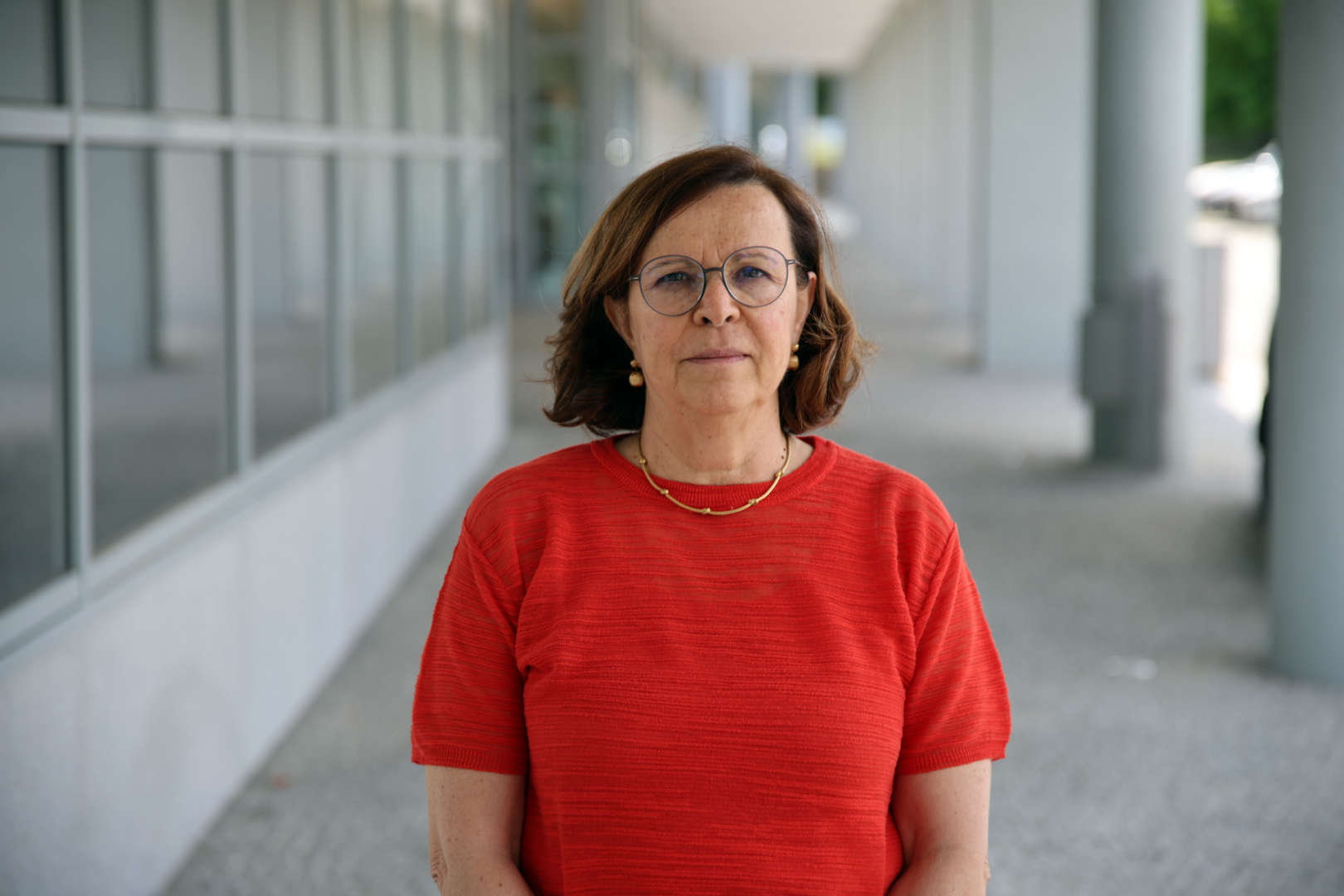
O meu nome é Ana Maria Mendonça e sou Professora Associada do Departamento de Engenharia Eletrotécnica (DEEC) da Faculdade de Engenharia da Universidade do Porto (FEUP). Foi nesta Universidade que concluí o meu doutoramento em 1994. Fui investigadora do Instituto de Engenharia Biomédica (INEB) até 2014, mas a partir de 2015 integrei o Centro de Investigação em Engenharia Biomédica em do INESC TEC como investigadora sénior.

Na minha atividade de gestão de ensino superior e investigação, fui membro do Conselho Executivo do DEEC e, mais recentemente, Subdiretora da FEUP. No INEB, integrei a Direção do Instituto inicialmente como vogal e, posteriormente, como Presidente da Direção.

Fui membro eleito do Conselho Científico da FEUP e sou atualmente membro do Conselho Pedagógico desta escola. Integrei as comissões científicas de vários ciclos de estudo da FEUP e sou atualment Diretora da Licenciatura e do Mestrado em BioEngenharia, do Mestrado em Engenharia Biomédica e do Programa Doutoral em Engenharia Biomédica da FEUP.

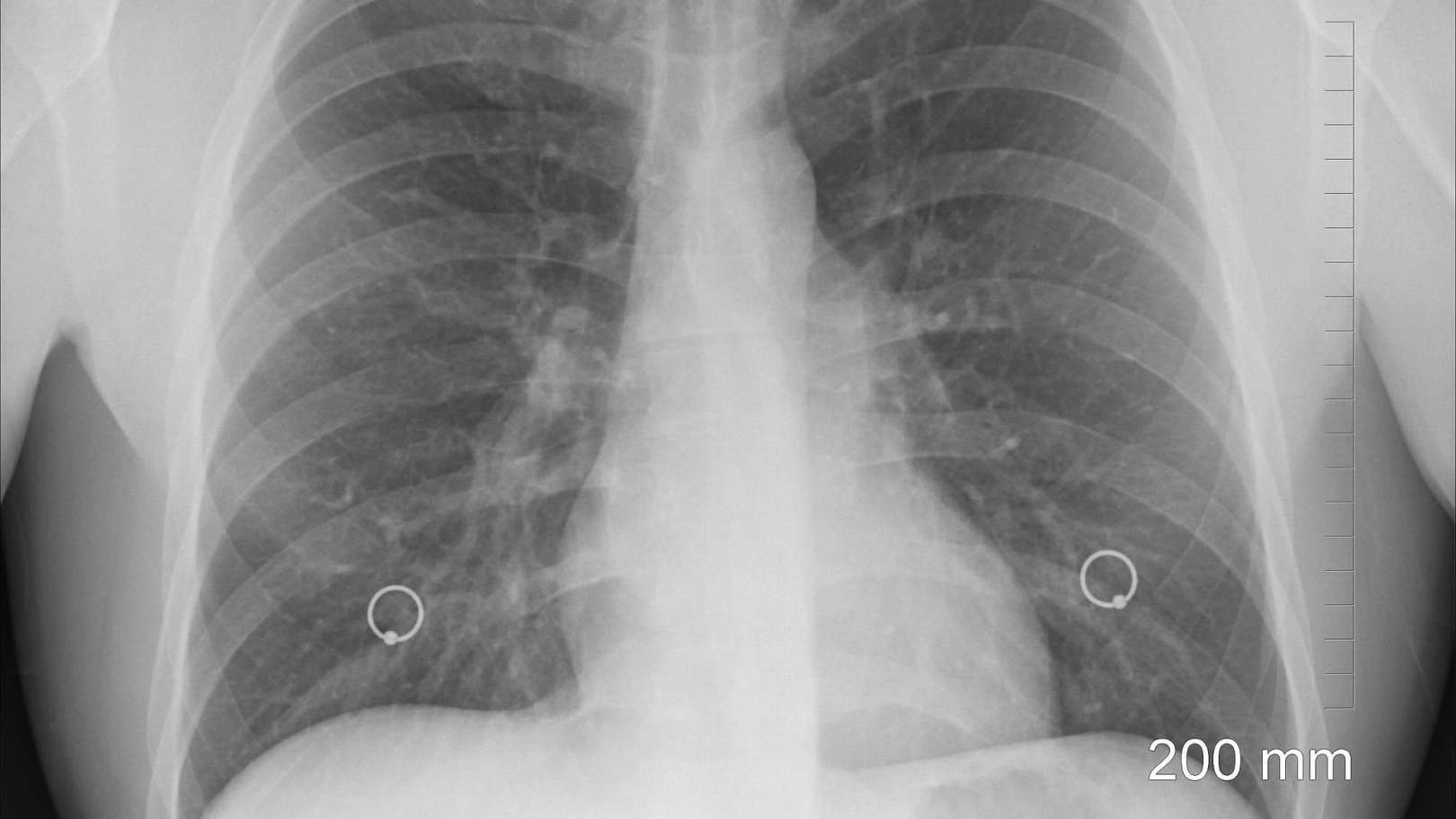
Tenho colaborado como investigadora ou como responsável em diversos projetos de investigação, principalmente na área da imagem biomédica. O meu trabalho de investigação centrou-se essencialmente no desenvolvimento de metodologias de análise de imagem e classificação tendo como objetivo a extração de informação útil de imagens médicas para apoiar o diagnóstico médico. O trabalho passado foi dedicado essencialmente às patologias da retina, do pulmão e doenças genéticas, mas o trabalho atual está essencialmente focado no desenvolvimento de sistema de apoio ao diagnóstico em oftalmologia e radiologia.

Tópicos
de interesse
Detalhes

Detalhes

  • Nome

    Ana Maria Mendonça
  • Cargo

    Investigador Sénior
  • Desde

    01 janeiro 2015
  • Nacionalidade

    Portugal
  • Contactos

    +351222094106
    ana.mendonca@inesctec.pt
005
Publicações

2024

STERN: Attention-driven Spatial Transformer Network for abnormality detection in chest X-ray images

Autores
Rocha, J; Pereira, SC; Pedrosa, J; Campilho, A; Mendonça, AM;

Publicação
ARTIFICIAL INTELLIGENCE IN MEDICINE

Abstract
Chest X-ray scans are frequently requested to detect the presence of abnormalities, due to their low-cost and non-invasive nature. The interpretation of these images can be automated to prioritize more urgent exams through deep learning models, but the presence of image artifacts, e.g. lettering, often generates a harmful bias in the classifiers and an increase of false positive results. Consequently, healthcare would benefit from a system that selects the thoracic region of interest prior to deciding whether an image is possibly pathologic. The current work tackles this binary classification exercise, in which an image is either normal or abnormal, using an attention-driven and spatially unsupervised Spatial Transformer Network (STERN), that takes advantage of a novel domain-specific loss to better frame the region of interest. Unlike the state of the art, in which this type of networks is usually employed for image alignment, this work proposes a spatial transformer module that is used specifically for attention, as an alternative to the standard object detection models that typically precede the classifier to crop out the region of interest. In sum, the proposed end-to-end architecture dynamically scales and aligns the input images to maximize the classifier's performance, by selecting the thorax with translation and non-isotropic scaling transformations, and thus eliminating artifacts. Additionally, this paper provides an extensive and objective analysis of the selected regions of interest, by proposing a set of mathematical evaluation metrics. The results indicate that the STERN achieves similar results to using YOLO-cropped images, with reduced computational cost and without the need for localization labels. More specifically, the system is able to distinguish abnormal frontal images from the CheXpert dataset, with a mean AUC of 85.67% -a 2.55% improvement vs. the 0.98% improvement achieved by the YOLO-based counterpart in comparison to a standard baseline classifier. At the same time, the STERN approach requires less than 2/3 of the training parameters, while increasing the inference time per batch in less than 2 ms. Code available via GitHub.

2024

Automated image label extraction from radiology reports - A review

Autores
Pereira, SC; Mendonca, AM; Campilho, A; Sousa, P; Lopes, CT;

Publicação
ARTIFICIAL INTELLIGENCE IN MEDICINE

Abstract
Machine Learning models need large amounts of annotated data for training. In the field of medical imaging, labeled data is especially difficult to obtain because the annotations have to be performed by qualified physicians. Natural Language Processing (NLP) tools can be applied to radiology reports to extract labels for medical images automatically. Compared to manual labeling, this approach requires smaller annotation efforts and can therefore facilitate the creation of labeled medical image data sets. In this article, we summarize the literature on this topic spanning from 2013 to 2023, starting with a meta-analysis of the included articles, followed by a qualitative and quantitative systematization of the results. Overall, we found four types of studies on the extraction of labels from radiology reports: those describing systems based on symbolic NLP, statistical NLP, neural NLP, and those describing systems combining or comparing two or more of the latter. Despite the large variety of existing approaches, there is still room for further improvement. This work can contribute to the development of new techniques or the improvement of existing ones.

2024

Human versus Artificial Intelligence: Validation of a Deep Learning Model for Retinal Layer and Fluid Segmentation in Optical Coherence Tomography Images from Patients with Age-Related Macular Degeneration

Autores
Miranda, M; Santos-Oliveira, J; Mendonca, AM; Sousa, V; Melo, T; Carneiro, A;

Publicação
DIAGNOSTICS

Abstract
Artificial intelligence (AI) models have received considerable attention in recent years for their ability to identify optical coherence tomography (OCT) biomarkers with clinical diagnostic potential and predict disease progression. This study aims to externally validate a deep learning (DL) algorithm by comparing its segmentation of retinal layers and fluid with a gold-standard method for manually adjusting the automatic segmentation of the Heidelberg Spectralis HRA + OCT software Version 6.16.8.0. A total of sixty OCT images of healthy subjects and patients with intermediate and exudative age-related macular degeneration (AMD) were included. A quantitative analysis of the retinal thickness and fluid area was performed, and the discrepancy between these methods was investigated. The results showed a moderate-to-strong correlation between the metrics extracted by both software types, in all the groups, and an overall near-perfect area overlap was observed, except for in the inner segment ellipsoid (ISE) layer. The DL system detected a significant difference in the outer retinal thickness across disease stages and accurately identified fluid in exudative cases. In more diseased eyes, there was significantly more disagreement between these methods. This DL system appears to be a reliable method for accessing important OCT biomarkers in AMD. However, further accuracy testing should be conducted to confirm its validity in real-world settings to ultimately aid ophthalmologists in OCT imaging management and guide timely treatment approaches.

2024

Distribution-based detection of radiographic changes in pneumonia patterns: A COVID-19 case study

Autores
Pereira, SC; Rocha, J; Campilho, A; Mendonça, AM;

Publicação
HELIYON

Abstract
Although the classification of chest radiographs has long been an extensively researched topic, interest increased significantly with the onset of the COVID-19 pandemic. Existing results are promising; however, the radiological similarities between COVID-19 and other types of respiratory diseases limit the success of conventional image classification approaches that focus on single instances. This study proposes a novel perspective that conceptualizes COVID-19 pneumonia as a deviation from a normative distribution of typical pneumonia patterns. Using a population- based approach, our approach utilizes distributional anomaly detection. This method diverges from traditional instance-wise approaches by focusing on sets of scans instead of individual images. Using an autoencoder to extract feature representations, we present instance-based and distribution-based assessments of the separability between COVID-positive and COVIDnegative pneumonia radiographs. The results demonstrate that the proposed distribution-based methodology outperforms conventional instance-based techniques in identifying radiographic changes associated with COVID-positive cases. This underscores its potential as an early warning system capable of detecting significant distributional shifts in radiographic data. By continuously monitoring these changes, this approach offers a mechanism for early identification of emerging health trends, potentially signaling the onset of new pandemics and enabling prompt public health responses.

2024

Evaluating Visual Explainability in Chest X-Ray Pathology Detection

Autores
Pereira, P; Rocha, J; Pedrosa, J; Mendonça, AM;

Publicação
2024 IEEE 22ND MEDITERRANEAN ELECTROTECHNICAL CONFERENCE, MELECON 2024

Abstract
Chest X-Ray (CXR), plays a vital role in diagnosing lung and heart conditions, but the high demand for CXR examinations poses challenges for radiologists. Automatic support systems can ease this burden by assisting radiologists in the image analysis process. While Deep Learning models have shown promise in this task, concerns persist regarding their complexity and decision-making opacity. To address this, various visual explanation techniques have been developed to elucidate the model reasoning, some of which have received significant attention in literature and are widely used such as GradCAM. However, it is unclear how different explanations methods perform and how to quantitatively measure their performance, as well as how that performance may be dependent on the model architecture used and the dataset characteristics. In this work, two widely used deep classification networks - DenseNet121 and ResNet50 - are trained for multi-pathology classification on CXR and visual explanations are then generated using GradCAM, GradCAM++, EigenGrad-CAM, Saliency maps, LRP and DeepLift. These explanations methods are then compared with radiologist annotations using previously proposed explainability evaluations metrics - intersection over union and hit rate. Furthermore, a novel method to convey visual explanations in the form of radiological written reports is proposed, allowing for a clinically-oriented explainability evaluation metric - zones score. It is shown that Grad-CAM++ and Saliency methods offer the most accurate explanations and that the effectiveness of visual explanations is found to vary based on the model and corresponding input size. Additionally, the explainability performance across different CXR datasets is evaluated, highlighting that the explanation quality depends on the dataset's characteristics and annotations.

Teses
supervisionadas

2024

XAIPrivacy – XAI with differential privacy

Autor
Fábio Araújo

Instituição
UP-FEUP

2024

Segmentation and Characterization of the Vascular Network in OCTA images

Autor
Matilde Carvalho Costa

Instituição

2024

Chest Radiography Content-Based Image Retrieval

Autor
Francisca Silva

Instituição
UP-FEUP

2023

Explainable Automatic Chest Radiography Pathology Detection and Reporting

Autor
Pedro Daniel Couto Pereira

Instituição
UP-FEUP

2023

Artificial Intelligence-based Decision Support Models for COVID-19 Detection

Autor
Sofia Perestrelo de Vasconcelos Cardoso Pereira

Instituição
UP-FEUP