Cookies
O website necessita de alguns cookies e outros recursos semelhantes para funcionar. Caso o permita, o INESC TEC irá utilizar cookies para recolher dados sobre as suas visitas, contribuindo, assim, para estatísticas agregadas que permitem melhorar o nosso serviço. Ver mais
Aceitar Rejeitar
  • Menu
Sobre

Sobre

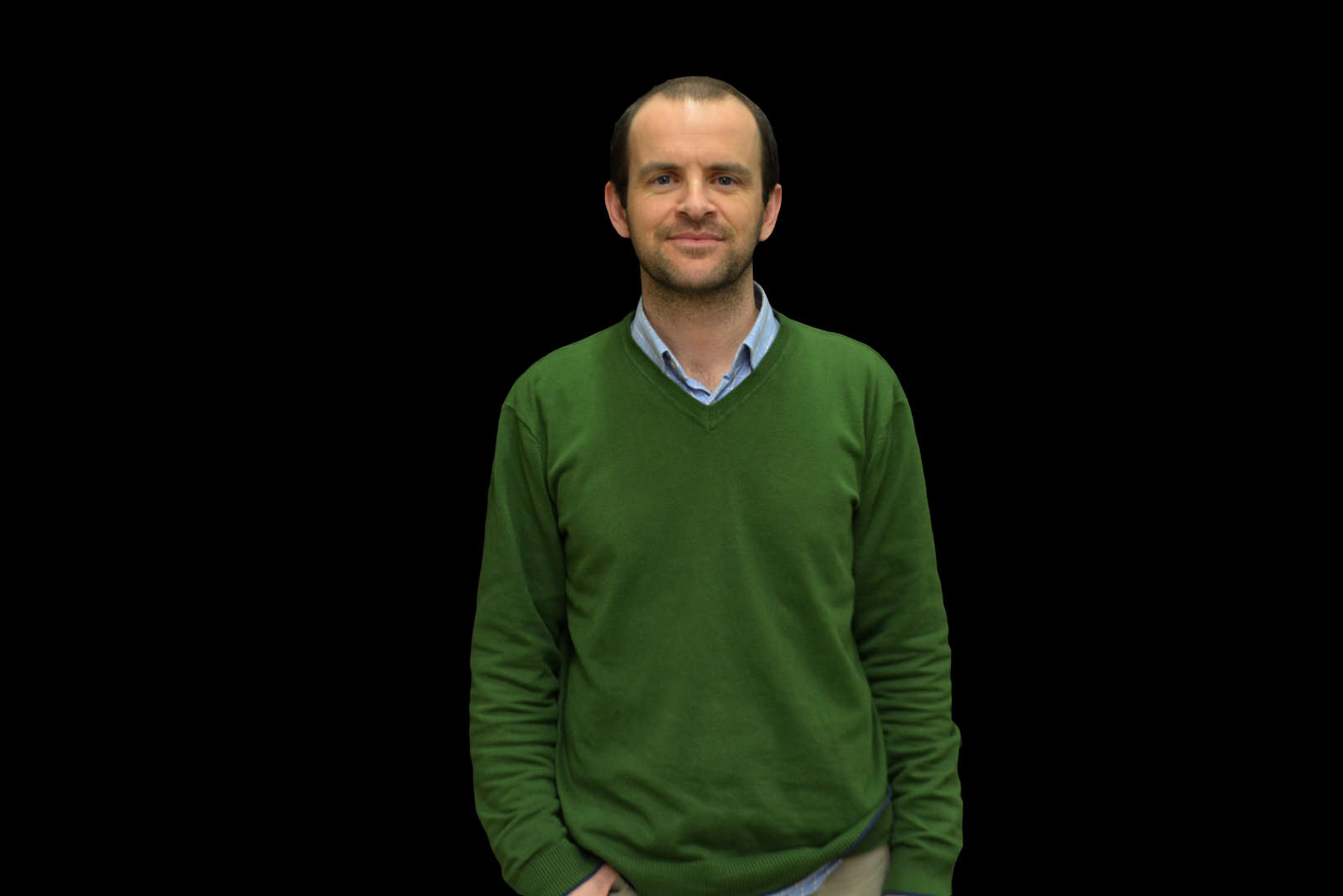
Jaime S. Cardoso, licenciado em Engenharia e Eletrotécnica e de Computadores em 1999, Mestre em Engenharia Matemática em 2005 e doutorado em Visão Computacional em 2006, todos pela Universidade do Porto. Professor Associado com agregação na Faculdade de Engenharia da Universidade do Porto (FEUP) e Investigador Sénior em 'Information Processing and Pattern Recognition' no Centro de Telecomunicações e Multimédia do INESC TEC.

A sua investigação assenta em três grandes domínios: visão computacional, "machine learning" e sistemas de suporte à decisão. A investigação em processamento de imagem e vídeo tem abordado a área de biometria, imagem médica e "video tracking" para aplicações de vigilância e desportos. O trabalho em "machine learning" foca-se na adaptação de sistemas de aprendizagem às condições desafiantes de informação visual. A ênfase dos sistemas de suporte à decisão tem sido dirigida a aplicações médicas, sempre ancoradas com a análise automática de informação visual.

É co-autor de mais de 150 artigos, dos quais mais de 50 em jornais internacionais, com mais de 6500 citações (google scholar). Foi investigador principal em 6 projectos de I&D e participou em 14 projectos de I&D, incluindo 5 projectos europeus e um contrato directo com a BBC do Reino Unido.

Tópicos
de interesse
Detalhes

Detalhes

  • Nome

    Jaime Cardoso
  • Cargo

    Investigador Coordenador
  • Desde

    15 setembro 1998
019
Publicações

2025

A survey on cell nuclei instance segmentation and classification: Leveraging context and attention

Autores
Nunes, JD; Montezuma, D; Oliveira, D; Pereira, T; Cardoso, JS;

Publicação
MEDICAL IMAGE ANALYSIS

Abstract
Nuclear-derived morphological features and biomarkers provide relevant insights regarding the tumour microenvironment, while also allowing diagnosis and prognosis in specific cancer types. However, manually annotating nuclei from the gigapixel Haematoxylin and Eosin (H&E)-stained Whole Slide Images (WSIs) is a laborious and costly task, meaning automated algorithms for cell nuclei instance segmentation and classification could alleviate the workload of pathologists and clinical researchers and at the same time facilitate the automatic extraction of clinically interpretable features for artificial intelligence (AI) tools. But due to high intra- and inter-class variability of nuclei morphological and chromatic features, as well as H&Estains susceptibility to artefacts, state-of-the-art algorithms cannot correctly detect and classify instances with the necessary performance. In this work, we hypothesize context and attention inductive biases in artificial neural networks (ANNs) could increase the performance and generalization of algorithms for cell nuclei instance segmentation and classification. To understand the advantages, use-cases, and limitations of context and attention-based mechanisms in instance segmentation and classification, we start by reviewing works in computer vision and medical imaging. We then conduct a thorough survey on context and attention methods for cell nuclei instance segmentation and classification from H&E-stained microscopy imaging, while providing a comprehensive discussion of the challenges being tackled with context and attention. Besides, we illustrate some limitations of current approaches and present ideas for future research. As a case study, we extend both a general (Mask-RCNN) and a customized (HoVer-Net) instance segmentation and classification methods with context- and attention-based mechanisms and perform a comparative analysis on a multicentre dataset for colon nuclei identification and counting. Although pathologists rely on context at multiple levels while paying attention to specific Regions of Interest (RoIs) when analysing and annotating WSIs, our findings suggest translating that domain knowledge into algorithm design is no trivial task, but to fully exploit these mechanisms in ANNs, the scientific understanding of these methods should first be addressed.

2025

CNN explanation methods for ordinal regression tasks

Autores
Gómez, JB; Cruz, RPM; Cardoso, JS; Gutiérrez, PA; Martínez, CH;

Publicação
Neurocomputing

Abstract

2025

CNN explanation methods for ordinal regression tasks

Autores
Barbero-Gómez, J; Cruz, RPM; Cardoso, JS; Gutiérrez, PA; Hervás-Martínez, C;

Publicação
NEUROCOMPUTING

Abstract
The use of Convolutional Neural Network (CNN) models for image classification tasks has gained significant popularity. However, the lack of interpretability in CNN models poses challenges for debugging and validation. To address this issue, various explanation methods have been developed to provide insights into CNN models. This paper focuses on the validity of these explanation methods for ordinal regression tasks, where the classes have a predefined order relationship. Different modifications are proposed for two explanation methods to exploit the ordinal relationships between classes: Grad-CAM based on Ordinal Binary Decomposition (GradOBDCAM) and Ordinal Information Bottleneck Analysis (OIBA). The performance of these modified methods is compared to existing popular alternatives. Experimental results demonstrate that GradOBD-CAM outperforms other methods in terms of interpretability for three out of four datasets, while OIBA achieves superior performance compared to IBA.

2024

Classification of Pulmonary Nodules in 2-[<SUP>18</SUP>F]FDG PET/CT Images with a 3D Convolutional Neural Network

Autores
Alves, VM; Cardoso, JD; Gama, J;

Publicação
NUCLEAR MEDICINE AND MOLECULAR IMAGING

Abstract
Purpose 2-[F-18]FDG PET/CT plays an important role in the management of pulmonary nodules. Convolutional neural networks (CNNs) automatically learn features from images and have the potential to improve the discrimination between malignant and benign pulmonary nodules. The purpose of this study was to develop and validate a CNN model for classification of pulmonary nodules from 2-[F-18]FDG PET images.Methods One hundred thirteen participants were retrospectively selected. One nodule per participant. The 2-[F-18]FDG PET images were preprocessed and annotated with the reference standard. The deep learning experiment entailed random data splitting in five sets. A test set was held out for evaluation of the final model. Four-fold cross-validation was performed from the remaining sets for training and evaluating a set of candidate models and for selecting the final model. Models of three types of 3D CNNs architectures were trained from random weight initialization (Stacked 3D CNN, VGG-like and Inception-v2-like models) both in original and augmented datasets. Transfer learning, from ImageNet with ResNet-50, was also used.Results The final model (Stacked 3D CNN model) obtained an area under the ROC curve of 0.8385 (95% CI: 0.6455-1.0000) in the test set. The model had a sensibility of 80.00%, a specificity of 69.23% and an accuracy of 73.91%, in the test set, for an optimised decision threshold that assigns a higher cost to false negatives.Conclusion A 3D CNN model was effective at distinguishing benign from malignant pulmonary nodules in 2-[F-18]FDG PET images.

2024

Active Supervision: Human in the Loop

Autores
Cruz, RPM; Shihavuddin, ASM; Maruf, MH; Cardoso, JS;

Publicação
PROGRESS IN PATTERN RECOGNITION, IMAGE ANALYSIS, COMPUTER VISION, AND APPLICATIONS, CIARP 2023, PT I

Abstract
After the learning process, certain types of images may not be modeled correctly because they were not well represented in the training set. These failures can then be compensated for by collecting more images from the real-world and incorporating them into the learning process - an expensive process known as active learning. The proposed twist, called active supervision, uses the model itself to change the existing images in the direction where the boundary is less defined and requests feedback from the user on how the new image should be labeled. Experiments in the context of class imbalance show the technique is able to increase model performance in rare classes. Active human supervision helps provide crucial information to the model during training that the training set lacks.

Teses
supervisionadas

2023

Deep Learning for improved NCP quantification and false positive reduction in CCTA

Autor
Maria Carolina Morgado Bastião Guedes Brás

Instituição
UP-FEUP

2023

Annotation-Efficient Segmentation of Medical Images for Cancer Detection

Autor
Pedro Miguel Vendas da Costa

Instituição
UP-FEUP

2023

Drawing the Line: Multimodal Lane Estimation for Autonomous Vehicles

Autor
Cláudia Maria Eira Ribeiro

Instituição
UP-FEUP

2023

Application of Explainable AI in Deep Learning Models

Autor
Mariana Margalho Alves Calado

Instituição
UP-FEUP

2023

Interpretable Machine Learning and its Application to Medical Decision Support Systems

Autor
Tiago Filipe Sousa Gonçalves

Instituição
UP-FEUP